What Your Dental X-Rays Actually Show (And Why They’re Not Just About Cavities)
“My teeth feel completely fine — why do I need X-rays again this year?”
I get this question a lot, and it’s a fair one. If nothing hurts and nothing looks wrong in the mirror, X-rays can feel like an extra step tacked onto a routine visit rather than something genuinely necessary. The honest answer is that most of what X-rays actually catch has nothing to do with how your teeth feel — that’s precisely the point of taking them. Let me walk through what each type of X-ray actually shows, why they catch things a visual exam physically can’t, and what the radiation exposure actually amounts to, since that’s usually the real question underneath the first one.
The four types you’ll actually encounter
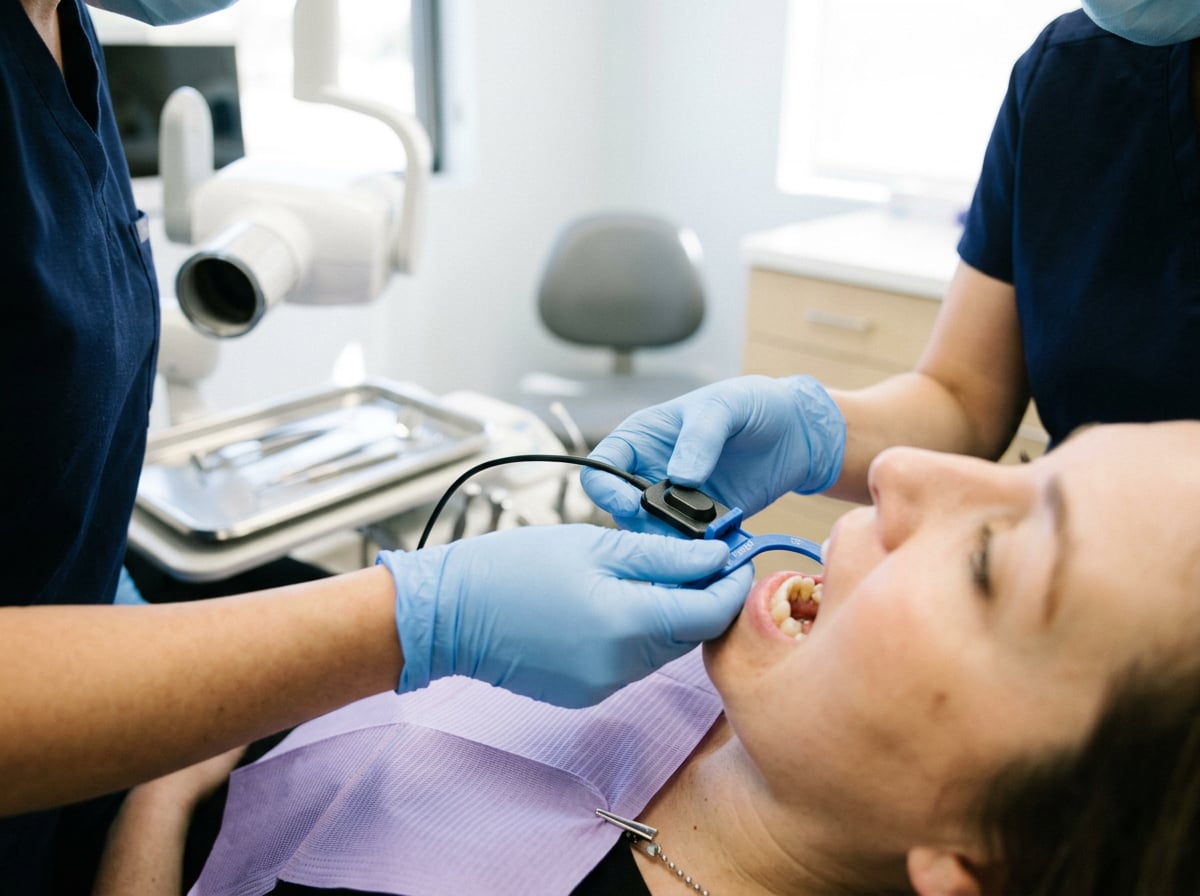
Bitewing X-rays are the ones you’re probably most familiar with — you bite down on a small wing-shaped tab while the image captures the crowns of your upper and lower teeth in one area of your mouth. These are the workhorse image for catching decay between teeth, which is invisible to a direct look, and for checking bone levels that indicate gum disease.
Periapical X-rays capture an entire tooth in a single image, from the crown all the way past the root tip to the bone anchoring it in your jaw. These are what we rely on for problems bitewings can’t show — an infection or abscess forming at the root tip, an issue with a tooth’s root structure, or bone changes around a specific tooth that need close, individual evaluation.
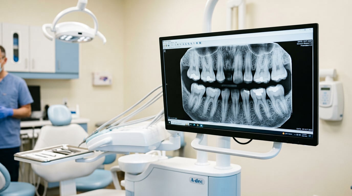
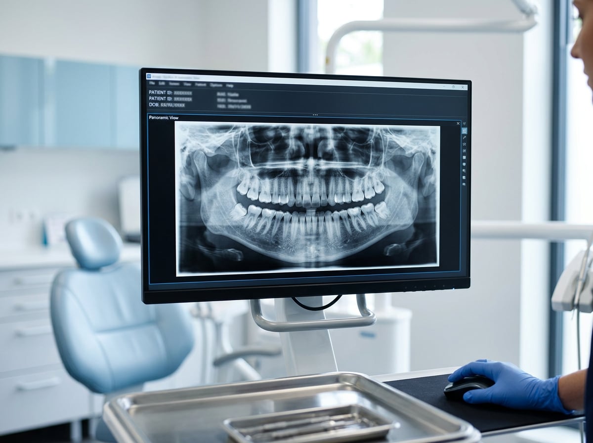
Panoramic X-rays capture your entire mouth in one wide image — both jaws, all your teeth, your sinuses, and the joints where your jaw connects to your skull. This is the image used to track wisdom teeth, spot impacted teeth, evaluate jaw development, or get an overview before planning something like an implant or extraction.
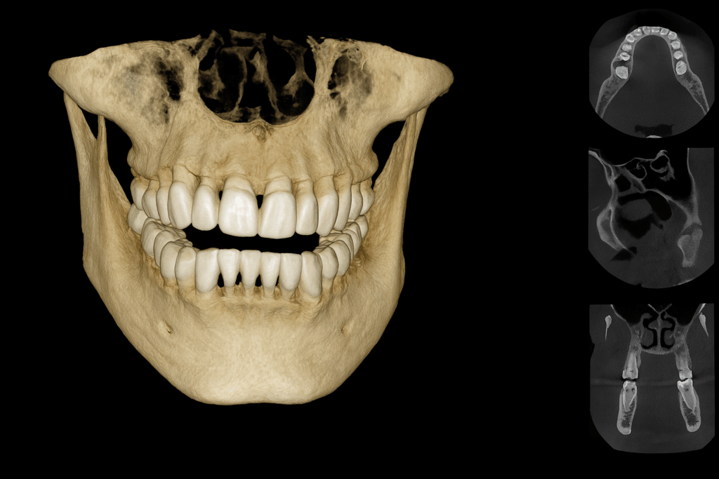
Cone-beam CT (CBCT) scans go a step further, building an actual 3D image of your jaw rather than a flat 2D picture. These are reserved for more complex planning — implant placement, evaluating bone volume before surgery, or investigating something a 2D image left ambiguous — rather than routine use. Because CBCT delivers a higher radiation dose than a standard 2D image, current guidance is specific that it should be reserved for cases where a 2D image genuinely isn’t enough, with the smallest field of view that still answers the clinical question, rather than used as a default upgrade.
What X-rays catch that a visual exam physically cannot
This is really the core of why X-rays matter regardless of how your mouth feels. A dentist looking directly at your teeth can only assess the surfaces actually visible to the eye. X-rays reveal the parts of the picture that visual inspection structurally cannot reach:
Decay between teeth. The contact point where two teeth touch is completely hidden from direct view, and this is one of the most common places cavities develop. Bitewing X-rays are often the only way this kind of decay gets caught before it’s large enough to be felt.
Bone loss from gum disease. Advanced gum disease erodes the bone supporting your teeth, and the rate and pattern of that loss is only reliably visible on an X-ray, not through a visual look at the gums alone.
Infections at the root tip. An abscess or infection brewing at the tip of a tooth’s root often causes no visible change to the tooth or surrounding gum in its early stages, and periapical X-rays are frequently how these are caught before they cause pain.
Impacted or developing teeth. Wisdom teeth that haven’t erupted, or a permanent tooth that’s developing in an unusual position in a younger patient, are entirely invisible without a panoramic image.
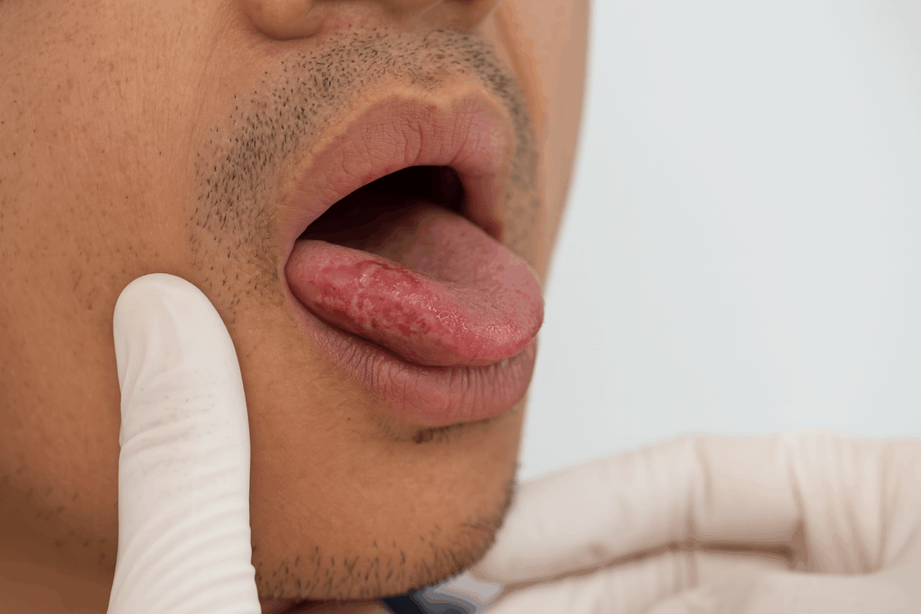
Cysts and other abnormalities in the jaw. Occasionally an X-ray reveals something entirely unrelated to a specific tooth — a cyst, a growth, or a structural abnormality in the jawbone — found incidentally during a routine image rather than because it was causing symptoms.
Did You Know
A full set of four bitewing X-rays exposes you to roughly the same radiation as spending a day or two in a typical brick or concrete home, and a single digital periapical X-ray is comparable to eating about 50 bananas’ worth of naturally occurring radiation. Dental X-rays are consistently among the lowest-dose imaging used in medicine.
Addressing the radiation question directly
This is almost always the real concern behind “do I actually need this X-ray,” so it’s worth being specific rather than just reassuring. Modern digital dental X-rays use a small fraction of the radiation older film-based systems required, and the American Dental Association has stated plainly that radiation exposure from dental X-rays is minimal compared to both background environmental radiation and other medical imaging. For context, a chest X-ray delivers roughly 20 to 100 times more radiation than a single digital dental image, and a CT scan of any kind delivers dramatically more than that. A full mouth series is measured in single-digit microsieverts — a fraction of the roughly 620 millirem the average American is exposed to annually just from natural background sources like soil, radon, and cosmic radiation. That doesn’t mean X-rays should be taken indiscriminately, and it isn’t the argument for that — it means the actual dose involved is not the reason to skip an X-ray your dentist has a real diagnostic reason to recommend.
How often you actually need them
This has genuinely shifted in recent guidance. Updated recommendations released by the American Dental Association in early 2026 emphasize that X-ray decisions should be based on an individual review of your medical and dental history, prior images, and current clinical exam findings — not a fixed schedule applied to everyone the same way. In practice, that means a patient with a low cavity risk and stable dental history might only need bitewings every 18 to 24 months, while a patient with a history of frequent decay, gum disease, or ongoing treatment might need them more often. Panoramic images are typically spaced much further apart — often years, not annually — unless something specific warrants a closer look sooner.
Worth Knowing
The 2026 ADA guidance specifically frames this as imaging being used “most effectively in moderation” — meaning both directions matter. Too few X-rays risks missing decay, bone loss, or infections early, while X-rays taken without a real clinical reason add exposure without adding diagnostic value. The right frequency is genuinely individual, not a blanket rule either way.
What actually gets missed if you skip X-rays for years
Decay between teeth progresses silently. Without bitewings, cavities forming at the contact points between teeth often aren’t caught until they’re large enough to be visible, painful, or both — well past the point where a small filling would have been the fix.
Bone loss from gum disease goes untracked. Without periodic imaging, there’s no objective way to know whether periodontal treatment is actually holding steady or whether bone loss is quietly continuing beneath gums that may not look dramatically different to the eye.
Root infections aren’t caught until they hurt. An abscess brewing at a root tip can exist for a meaningful stretch of time without pain, and by the time it’s symptomatic, treatment is often more involved than it would have been if caught on a routine periapical image.
Impacted teeth go unnoticed until they cause a problem. Wisdom teeth or other impacted teeth can shift, crowd neighboring teeth, or develop cysts around them without any symptoms at all until the situation has become more complex to manage.
Failing old dental work goes undetected underneath. A filling or crown can develop a small gap or recurrent decay underneath it that’s completely invisible from the surface, and periodic bitewings are frequently how this gets caught before the tooth underneath is significantly compromised.
X-rays aren’t just for finding problems — they’re for planning treatment too
It’s easy to think of X-rays purely as a detection tool, but a meaningful part of their value is in planning treatment that’s already been decided on. Before placing a dental implant, imaging shows the exact bone volume and density available, which determines whether a standard implant works as planned or whether additional steps are needed first. Before a root canal, a periapical X-ray maps the shape and length of the root canal system, which is essential information for treating it accurately. Even something as routine as fitting a crown benefits from an X-ray confirming there’s no hidden decay or infection underneath that a visual exam alone would miss. In these cases, the X-ray isn’t answering “is something wrong” — it’s answering “exactly what does this specific tooth or bone look like,” which is a different, and equally important, kind of information.
A few questions worth asking at your next visit
“What specifically are you looking for with this X-ray?” A reasonable question any time imaging is recommended, and a dentist following current guidance should have a specific answer tied to your history or exam findings, not just “it’s been a while.”
“Can you bring forward my previous X-rays for comparison?” Comparing a new image against an older one is often more diagnostically useful than either image alone, since it shows whether something has changed over time rather than just what a single snapshot looks like.
“Do I actually need the full series, or just specific views?” Depending on what’s being evaluated, a couple of targeted images sometimes answer the clinical question just as well as a larger set, and it’s a reasonable thing to ask about.
A note on pregnancy and X-ray safety
This comes up often enough to address directly: dental X-rays are considered safe during pregnancy when needed, particularly with the lead apron and thyroid collar used as standard protective measures, and the radiation dose involved is extremely low to begin with. That said, most dentists will postpone non-urgent, routine imaging until after delivery simply out of an abundance of caution, while still taking any X-ray that’s genuinely needed to diagnose or treat an active problem, since untreated infection or pain carries its own risks during pregnancy. If you’re pregnant and due for a routine X-ray, that’s worth a quick conversation rather than an automatic yes or no either way.
| Type | What it shows | Typical frequency | Best for |
|---|---|---|---|
| Bitewing | Crowns of upper & lower teeth in one area | Every 12–24 months, based on risk Most common |
Decay between teeth, bone level checks |
| Periapical | One full tooth, crown to root tip | As needed for a specific tooth | Root infections, abscesses, root structure issues |
| Panoramic | Entire mouth, both jaws, sinuses, joints | Every several years, or as needed | Wisdom teeth, impacted teeth, overall planning |
| CBCT (3D) | Full 3D structure of the jaw | Only when specifically indicated Case dependent |
Implant planning, complex surgical cases |
Which type of X-ray, and how often
X-rays aren’t a formality tacked onto your checkup — they’re often the only way to catch decay between teeth, bone loss from gum disease, or an infection at a root tip before any of it causes pain. The radiation involved is genuinely minimal, well below what most patients assume, and the actual question worth asking isn’t “should I avoid X-rays” but “does my dentist have a specific reason for this one, based on my own history.” Modern guidance is explicit that imaging should be individualized rather than automatic, which means it’s entirely reasonable to ask what a specific image is for. If you’re ever unsure why a particular X-ray is being recommended, that’s a completely reasonable thing to ask before saying yes — and a dentist following current best practice should be glad to answer it.
Washington, PA & Pittsburgh
Wondering what your X-rays would actually catch?
Dr. Wakim can review your history and tell you honestly which imaging you actually need — not a one-size-fits-all schedule.

Dr. Elizabeth Wakim, DDS, is the founder of Enhanced Wellness. She’s a compassionate and highly-regarded dentist with her own practice in Washington, Pennsylvania, known for providing modern, comprehensive dental care, botox and facial aesthetics with a focus on patient comfort and anxiety reduction, serving general, cosmetic, and pediatric dentistry needs.